General Microbiology
Agar and Broth
The oCelloScope is used in microbiology, for live-cell imaging of bacteria or fungi in solution and on agar. You can use your micro titer plate of choice as long as it comes with a flat bottom. For agar work we recommend using our BioSense Solutions mini agar disks, developed for 12 well plates. Setting up experiments in the UniExplorer software is easy and current applications include: AMR Discovery, Preservatives Testing, Drug Efficacy Testing, Spike Testing, Medium Optimization, Probiotics Discovery, Co-Culture Studies and dedicated morphology analysis.
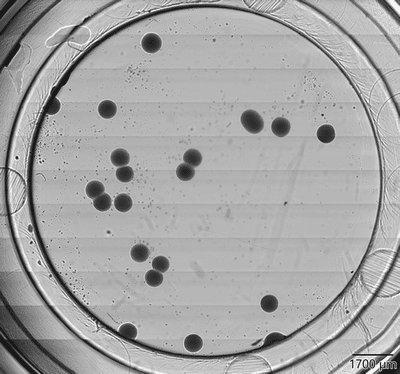
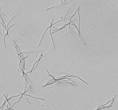
Left: Bacteria colonies merging on BioSense Solutions mini agar disk. Right: Enterobacter cloacae filamentation in solution
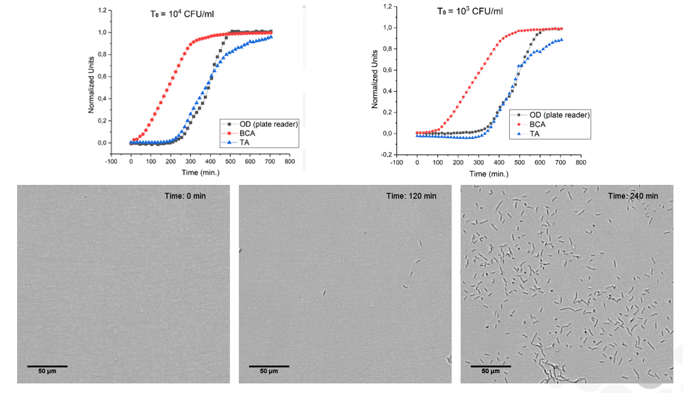
oCelloScope Vs. Plate reader
Measuring pixel intensities is approximately 250 times more sensitive than using OD600. With the BCA (Background Corrected Absorbance) algorithm we detect growth from 10_3 cells/ml. In curve to the right E-coli is inoculated in a 96-well and growth detected over time. Red curve is the BCA algorithm, while blue is an algorithm (TA) mimicking OD600. Black curve is the Varioscan plate reader. For validation images from well is shown. Inoculation is done at 10_3. The oCelloScope starts detecting at 5x10_3 and growth is detected at around 100 minutes. Notice that after 240 minutes you see many bacteria in image but no detection using OD600.
Medium Optimization
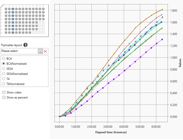
The oCelloScope is routinely used with medium manufacturers for optimization of nutrients in medium. Experiments can be done in high throughput and results easily visualized in the software.
Inoculate 100µl bacterial sample in a 96-well plate. Typical concentration used is 10_5 – 10_6. Program oCelloScope using the growth kinetic module and time interval of your choice. For log phase bacteria you would image every 30 minutes and for fungal spores every hour or two.
Data can be exported as excel or csv for you to analyze data externally.
Tracking colonies on agar
In microbiology it is desired to compare bacterial or fungal growth on agar with what you establish in solution. Applications, span from drug response, biopesticides, preservatives testing to basic research on bacterial morphology and growth speeds.
BioSense Solutions have developed a unique tracking module for analyzing growth on agar. The oCelloScope is able to count and track colonies from just 10µm in size. Comparing to standard agar plating in agar dishes we both visualize and count more colonies compared to a classical visual agar plate inspection. Reason is that many CFU merge as micro colonies and therefore counted as one in traditional agar counts.
We recommend using the BioSense mini agar disks, fitted to 12-well plates. Pipette approximately 170µl warm agar into a mini agar disk. Let disks solidify and inoculate 3-5µl sample on top. Use a microbiology loop to spread sample. Let disks rest for 15 minutes and then place disks upside down in micro titer plate. In the software, choose full well function and the mini disk plate layout. Set focus manually, by locating the surface. If bacteria are sub-micron it is advised to add beads to sample for you to better localize the surface. The oCelloScope will image the surface of disks and track growth of bacteria/fungi from 10µm in size.
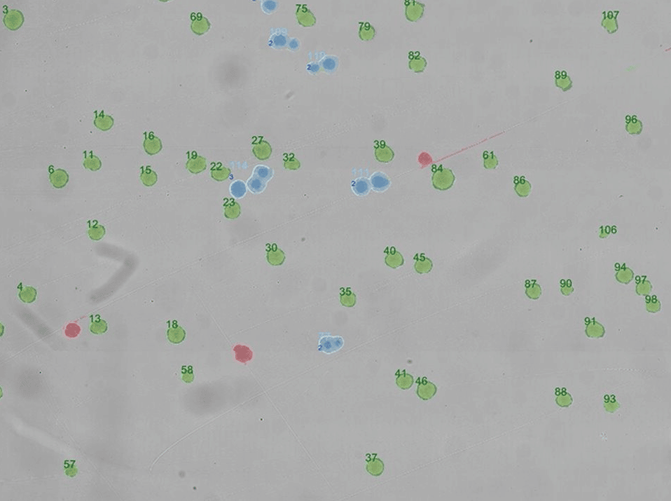
On the top right is a small area of a mini agar disk. Growing bacteria is counted and tracked over time. If colonies merge (blue), they are deleted from analysis. Objects not tracked are red.
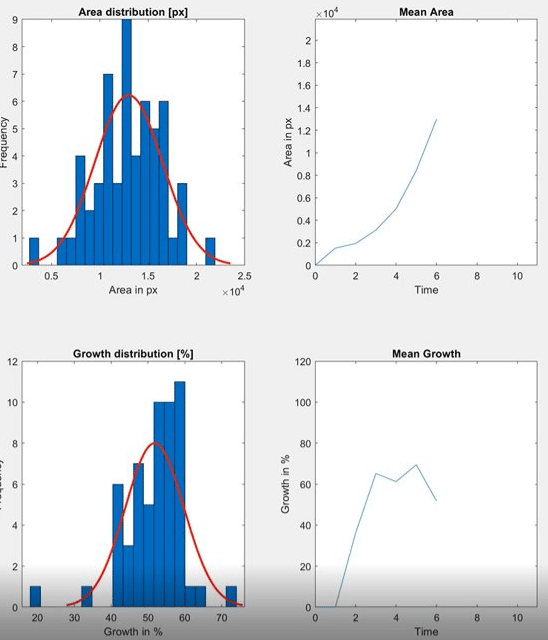
On the lower right is examples of data extracted from analysis (area distribution, growth distribution and means thereof).

Counting and tracking fungal spores
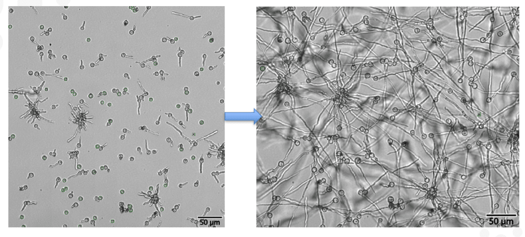
With the use of the counting module in the UniExplorer V12 software you can count and track fungal spores over time. The algorithm will find all specific non-germinated spores throughout your experiment. Once spores develop a germ tube, algorithms are designed to let go. The unique counting module will enable you to perform fast counts in high throughput and quantify germination.
The lab workhorse
With the size of an A4 sheet of paper, the oCelloScope might just have the smallest footprint of any instrument in your microbiology lab. The unique imaging technology, dedicated algorithms and easy interface might also make it the busiest instrument in your lab. “I wish I had this instrument during my PhD.”, is the most common remark we get.
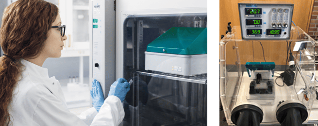
The right shows the oCelloScope inside an incubator and inside an anaerobic chamber.
Ready to get started?
We are ready to help you solve your tough research challenges. Our highly qualified team can help advance your next big discovery.
