Real-time monitoring and analyses of your fungi
oCelloScope is an automated live-cell imaging system that speeds up microbiology research and gives a deeper understanding of the biological processes.
Using standard microtiter plates, you can analyse up to 96 microbial samples at a time, and perform real-time analysis inside the incubator, saving hours of valuable time while maintaining the quality of your experiments.
Real-time monitoring of Aspergillus niger growth in malt extract broth (MEB) at 35°C over 17 hours.
Real-time analysis of Penicillium roqueforti growth with the SESA-Fungi algorithm.
Automated monitoring and analysis of fungal growth
Automated live monitoring and analysis of fungal growth is now possible with oCelloScope. OD is unreliable in measuring fungal growth, due to the morphology of the fungi and its non-uniform distribution. oCelloScope solves this issue by combining high resolution images with automated image analysis.
The SESA-Fungi algorithm, identifies the fungi in the image and quantifies the growth from spores to hyphae. Aunsbjerg SD et al. (2015), have shown that with oCelloScope results are obtained faster, compared to OD, resulting in a shorter incubation time.
Perform single cell analysis and follow the morphogenesis of your hyphae
With the build-in segmentation algorithms, the UniExplorer software enables the user to automatically perform single cell analysis, allowing scientists to follow the morphogenesis of single fungi cells, and discriminate and characterize the dynamics of different cell types in a complex sample.
More than 20 morphological features, including cell size and shape factors of each single object can be calculated and visualized in scatter plots and histograms, enabling the user to group the desired objects and analyse the morphology changes over time of the desired group.
As an example, discrimination between vegetative cells and spores in fungi or bacterial sporulation experiments can be performed

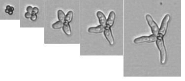
P.roqueforti spore germination time-lapse.
Real-time analysis of Penicillium roqueforti growth with the SESA-Fungi algorithm.
Automated monitoring and analysis of fungal growth
Automated live monitoring and analysis of fungal growth is now possible with oCelloScope. OD is unreliable in measuring fungal growth, due to the morphology of the fungi and its non-uniform distribution. oCelloScope solves this issue by combining high resolution images with automated image analysis.
The SESA-Fungi algorithm, identifies the fungi in the image and quantifies the growth from spores to hyphae. Aunsbjerg SD et al. (2015), have shown that with oCelloScope results are obtained faster, compared to OD, resulting in a shorter incubation time.

