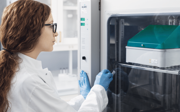
oCelloScope
oCelloScope – Automated Microbial Live-Cell Imaging and Analysis
Speed
- 250 times more sensitive than a plate reader (OD)
- Measure and visualize down to 5 x 10³ CFU/ml
- Growth kinetic MIC results in few hours Vs 16-20h using BMD
- FluidScope technology scans a full 96-well plate in less than 3 minutes
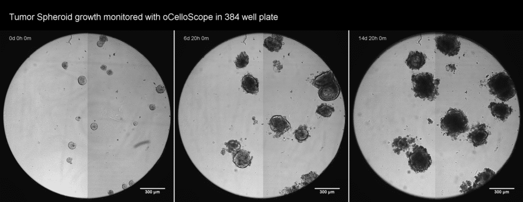
Early phase morphology
- Compare growth kinetic curves with images from each time-point
- Discover microorganism adaptation strategies
- Capture and quantify morphological changes over time
- Spheroplasts, Filamentation, Co-aggregation, Fungal spore germination
Value
- Full flexibility – use your standard microtiter plates
- You get full software package to use on multiple PC’s
- No expensive annual service contract
- Competitive pricing. Rent or Purchase – just ask for a quotation
Meet Our User Network

“A deep understanding of mechanisms of action of small molecules is critical for drug discovery. The research team at Venatorx Pharmaceuticals has incorporated oCelloScope, the state-of-the-art automated microscope developed by BioSense Solutions, to obtain valuable information on antibiotic effects on bacterial growth and morphology for the development of new antibiotics against clinically-important pathogens.”
Tsuyoshi Uehara
Principal Scientist of Biology, Venatorx Pharmaceuticals

“My laboratory has been using an oCelloScope™ to rapidly assess the effectiveness of various formulations for use in cleaning and sanitizing processing surfaces in the food industry. The lower detection limit and the ability to monitor cellular morphology make this instrument superior to using a microtiter plate reader to monitor media turbidity. The software is straight forward and easy to use. It has been a great laboratory tool.”
Lynne McLandsborough
Professor, and Department Head
Department of Food Science, University of Massachusetts
“Plate and biochemical assays can show strain and hit performances, but seeing is believing. The High Throughput Assay Development Team at Ginkgo Bioworks has incorporated the oCelloscope as an essential tool for validating and troubleshooting many of our assays. The oCelloscope reliably produces high-quality microscopy data from plate screens, building the scale of Ginkgo’s platform to engineer living cells to generate better products. Customer and inter-team understanding of strain growth, treatments, and communication of assay conditions and performances is made easy with the oCelloscope.”
Dr. Sanjiv Shah
Senior Test Engineer, Ginkgo Bioworks
“By using the oCelloScope we were able to follow germination of thousands of individual fungal conidiospores within a population or community. We could track development of these individuals by tracing size and shape and determine the influence of different conditions on their outgrowth. That was never done before”
Dr. Wieke R. Teertstra
Researcher, Microbiology, Kruyt building, Utrecht University

“The phage therapy group at University of Helsinki and Helsinki University Central Hospital has a long experience in conducting phagoram screenings in liquid culture using traditional adsorbance-based plate readers. The use of oCelloScope greatly increases the sensitivity of the measurement and makes it possible to conduct the whole phagoram assay within one working day.“
Dr. Saija Kiljunen // MSc. Sheetal Patpatia
PhD, Senior Scientist // MSc, Post-graduate student
Do you know how your cells develop?
The oCelloScope provides deeper insight and more information about your cells with real-time kinetic data. oCelloScope automatically acquires and analyzes images in standard micro-titer plates. We free up researchers’ time, increases sample throughput, and delivers richer data.
oCelloScope is a unique high throughput live-cell imaging system for sensitive and detailed monitoring of biological growth and development.
- Unique proprietary scanning technology, FluidScopeTM
- Unique Image analysis software based on both pixel detection and deep learning algorithms.
Watch the video and see oCelloScope in action
oCelloScope: Rapid, sensitive real-time Antimicrobial Susceptibility Testing
How can you benefit from live-cell image analysis with oCelloScope?
By combining continuous real-time imaging and powerful image analysis software with oCelloScope:
- You will never miss a data point again
- Capture and profile time dependent events
- Analyze microbial growth and development
- Visualize and validate the results with images and videos
oCelloScope Workflow
With oCelloScope and the powerful UniExplorer software your research are supported day and night.
Make your experiment in any environment
- oCelloScope fits inside your Incubator or anaerobic chamber
- Compatible with standard microtiter plates, from 6 to 384 wells
- Combine with our new mini plate heaters to analyze from ambient to 55C
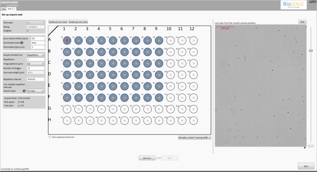
Setup automated acquisition and analysis
- Easy-to-use user interface for simple and flexible setup of your experiment
- Auto-focus and auto-illumination secures high quality data
Acquire images for as long as you like
- Automatically acquire images for hours, days or weeks
- Use UniExplorer’s continue mode for running multiple long-term experiments simultaneously
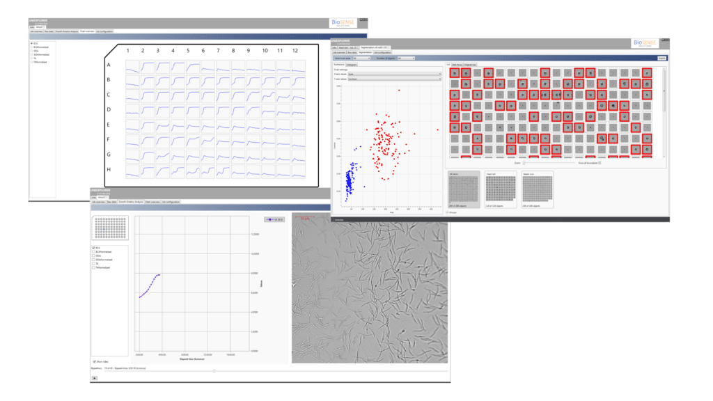
View and Analyze in real-time
- Follow you experiment in real-time with visualization of images and analysis
- Get a instant overview of your full 96-well plate
- Analyse single cells and mixed cultures
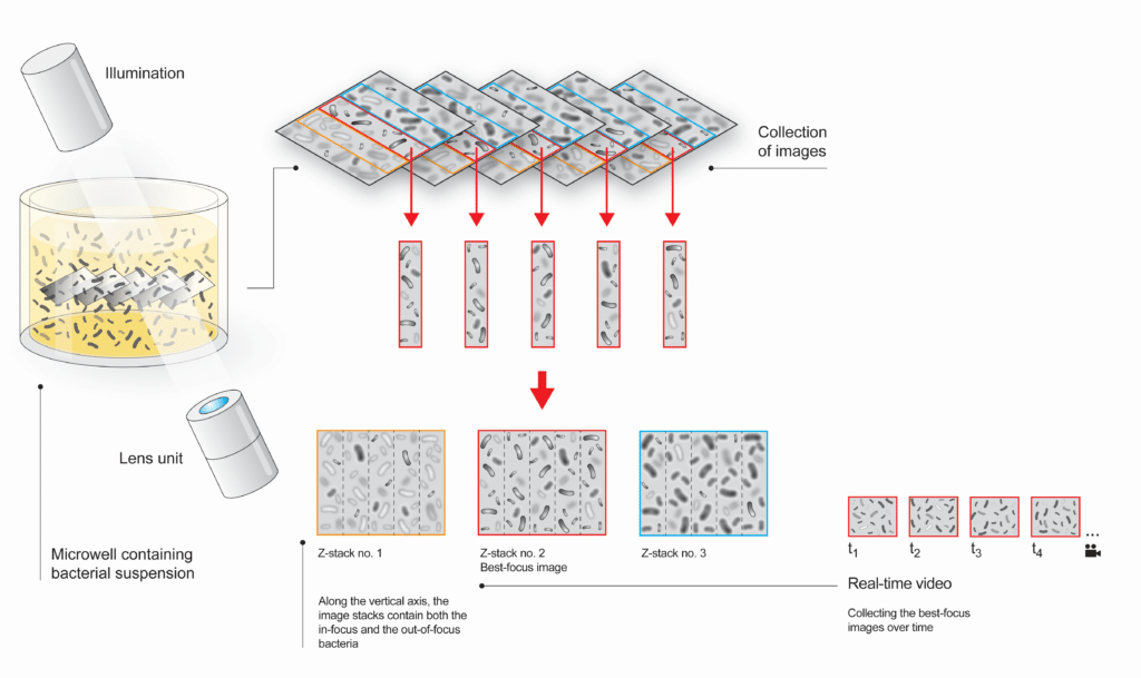
FluidScopeTM scanning technology
oCelloScope is based on a unique optical scanning technology FluidScopeTM combining optical techniques such as phase contrast, brightfield and confocal-like microscopy.
Together with advanced image processing algorithms, the FluidScopeTM technology provides:
- Fast scanning of a volume in the sample
- Greater freedom of operation with multiple Z-layers acquired simultaneously
- Detailed 3D information at a single cell level
- Requires no pre-treatment, staining or additional reagents
| Measurement specifications | |
|---|---|
| Sample objects |
Most objects in (semi-) transparent substances/liquid/agar can be analysed, e.g.:
|
| Sample matrix | Liquid and agar |
| Detectable density (sample concentration) | From 103 objects/mL |
| Detectable object size | 0.5 μm – 1 mm |
| Maximum scanning speed | 2 minutes 36 seconds (96 wells) |
| Optical magnification | 4x |
| Optical resolution | 1.3 µm |
| Camera | 5 Mpx, CCD, pixel size: 2,2 µm |
| Optical principle | FluidScopeTM (patent pending) |
| Environmental Specifications | |
| Operating temperature | 10 – 40°C |
| Operating humidity | 20 – 93 % RH |
| Dimensions | |
| Dimensions (D x W x H) | 450 x 260 x 250 mm (with open lid: H = 550 mm) |
| Weight | 9.6 kg |
| Connectors | RJ45 (Ethernet) |