Speed
- 250 times more sensitive than a plate reader (OD)
- Measure and visualize down to 5 x 10³ CFU/ml
- Growth kinetic MIC results in few hours Vs 16-20h using BMD
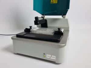
- FluidScope technology scans a full 96-well plate in less than 3 minutes
Early phase morphology
- Compare growth kinetic curves with images from each time-point
- Discover microorganism adaptation strategies
- Capture and quantify morphological changes over time
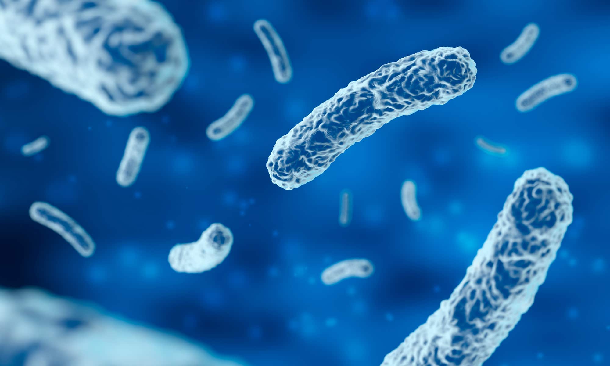
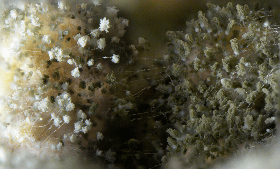
- Spheroplasts, Filamentation, Co-aggregation, Fungal spore germination
Value
- Full flexibility – use your standard microtiter plates
- You get full software package to use on multiple PC’s
- No expensive annual service contract
- Competitive pricing. Rent or Purchase – just ask for a quotation
Meet Our User Network

“The phage therapy group at University of Helsinki and Helsinki University Central Hospital has a long experience in conducting phagoram screenings in liquid culture using traditional adsorbance-based plate readers. The use of oCelloScope greatly increases the sensitivity of the measurement and makes it possible to conduct the whole phagoram assay within one working day.“

“By using the oCelloScope we were able to follow germination of thousands of individual fungal conidiospores within a population or community. We could track development of these individuals by tracing size and shape and determine the influence of different conditions on their outgrowth. That was never done before”

“Plate and biochemical assays can show strain and hit performances, but seeing is believing. The High Throughput Assay Development Team at Ginkgo Bioworks has incorporated the oCelloscope as an essential tool for validating and troubleshooting many of our assays. The oCelloscope reliably produces high-quality microscopy data from plate screens, building the scale of Ginkgo’s platform to engineer living cells to generate better products. Customer and inter-team understanding of strain growth, treatments, and communication of assay conditions and performances is made easy with the oCelloscope.”

Department of Food Science, University of Massachusetts
“My laboratory has been using an oCelloScope™ to rapidly assess the effectiveness of various formulations for use in cleaning and sanitizing processing surfaces in the food industry. The lower detection limit and the ability to monitor cellular morphology make this instrument superior to using a microtiter plate reader to monitor media turbidity. The software is straight forward and easy to use. It has been a great laboratory tool.”

“A deep understanding of mechanisms of action of small molecules is critical for drug discovery. The research team at Venatorx Pharmaceuticals has incorporated oCelloScope, the state-of-the-art automated microscope developed by BioSense Solutions, to obtain valuable information on antibiotic effects on bacterial growth and morphology for the development of new antibiotics against clinically-important pathogens.”
AMR – Antimicrobial Resistance
4 times faster than Broth Microdilution (BMD)
The oCelloScope is used by AMR researchers all over the world, including the US CDC to test new or existing drugs against resistant microorganisms.
..”All optical screening results were consistent with results from conventional susceptibility tests and were available in 4 h. This decreased the time required to determine antimicrobial susceptibility profiles by 75 to 80%, as conventional testing by BMD requires sufficient visible growth for interpretation of results and has been standardized by CLSI as a 16- to 20-h incubation time for B. anthracis.” McLaughlin, H.P., Gargis, A.S., Michel, P., Sue, D., and Weigel, L.M. (2017). J. Clin. Microbiol. 55, 959–970.
The software interface is intuitive and we typically only need a one hour web intro to get you started. Two modules are primarily used in AMR research – Growth Kinetics and Segmentation. Growth Kinetics is used to screen bacterial or fungal strains for resistance patterns in high throughput. The Segmentation module will take you a step deeper and enable you to analyze and quantify morphological responses.


Follow the β-lactam-induced cell morphology changes
The UniExplorer image analysis software enables you to get deeper insights into your sample dynamics, as changes on single cell level can be quantified.
Left: Segmentation analysis of Acinetobacter haemolyticus treated with Imipenem. Spheroplasts are detected using image analysis and counted automatically as a function of time.
Pull lever left or right to see the power of image analysis.
Food Microbiology

Fungal spore treatment and germination pattern (Aspergillus niger)
Time-lapse movie of Aspergillus niger
The oCelloScope for food microbiology enable you to analyze up to 96 microbial samples in real time. The higher sensitivity and the ability to monitor and analyze cellular morphology makes the oCelloScope superior to using a microplate reader. Today, researchers in Food Microbiology use the oCelloScope in: Preservatives Discovery, Screening of Probiotics, Sanitizer Development, Cheese Development, Yeast Discovery, Beverage Batch Release Testing, Food Molds and Fungal Spore Development.
Co-culture challenges in food microbiology
The UniExplorer image analysis software enables you to analyze Co-culture samples and discriminate between different species.
Right: Co-culture study of Lactobacillus and Yeast (Saccharomyces cerevisiae). Image analysis is used to measure inhibitory effects of Lactobacillus. Yeast growth morphological characteristics such as size, chain length and number of clusters can be monitored over time.
Pull lever left or right to see the power of image analysis.


oCelloScope Inspiration
Full well scanning of Escherichia coli. 2 µl inoculated on LB agar and development monitored in time-lapse. We scan a full 24 or 96 size well. Both in solution and on agar.
Pseudomonas aeruginosa growth time-lapse captured with oCelloScope: (left) non-treated control, (right) treated with 0.5µg/ml of Ceftazidime: Filamentation is observed as a consequence of antibiotic treatment. Starting cell concentration 105 cell/ml.
Use of oCelloScope flow-cell to monitor biofilm build up and dynamics
18h time-lapse of Pseudomonas aeruginosa challenged with 64mg/L Imipenem – far above MIC. Kinetics curve shows no growth as expected but note the formation of new 15-20um2 structures. Could these be persister cells?
Time-Lapse video showing stimulated T-Cells imaged every minute for five hours. Experiment was carried out in a customized T-flask.
Time-Lapse of PC12 Rat adrenal medulla cells. Field of view 0.6mm x 1.4mm.
Pharmaceutical research. Zebrafish, 72h embryo development. Dep of Veterinary and Animal Sciences, University of Copenhagen.
Fisheries Research. Ichtyophterius multifilis, Dep of Veterinary and Animal Sciences, University of Copenhagen.
Want to talk Science
or get a Quotation?
CALL US
Sales: (+45) 22 30 84 00
LOCATION
3520 Farum
Denmark